FUJIFILM and Department of Respiratory Medicine, Graduate School of Medicine, Kyoto University (Professor Toyohiro Hirai) have succeeded in jointly developing technology that uses artificial intelligence to automatically categorize and quantify lesions of interstitial pneumonia at an advanced level of precision. Fujifilm strives to introduce a diagnostic imaging support function that can incorporate this technology to its system solutions for clinical use starting from Japan by the end of fiscal year ending March 2021.
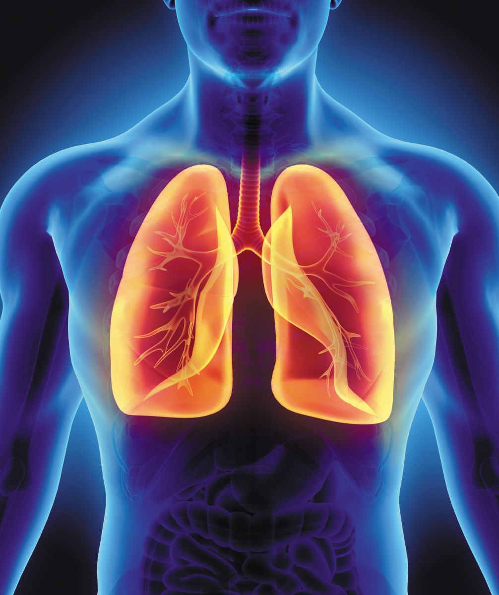
The lungs inspire oxygen and expire carbon dioxide through pulmonary alveoli. Interstitial pneumonia is a disease that develops when inflammation or damage to pulmonary alveolar wall causes interstitium tissue to thicken and harden. Interstitial pneumonia can be divided into those due to a clearly-defined cause, such as collagenosis like rheumatoid arthritis, pneumoconiosis, drug-induced pneumonia, and those without specific cause which is called idiopathic interstitial pneumonia (IIP).
IIP is a designated intractable disease that cannot be easily treated, and is classified into several types including idiopathic pulmonary fibrosis (IPF). Chest CT scan is one of the most useful examinations for diagnosis of IPF. However, CT images in IPF often show complex and various kinds of abnormal shadows, making it difficult to confirm the diagnosis especially at an early stage of the disease. In some cases, doctors visually observe the changes in individual lung lesions on CT images to confirm diagnosis and choose a treatment approach. In addition, it is known that IPF shows gradual changes in lesions on CT images as the disease progresses, and also sometimes a drastic change called “acute exacerbation” which leads to poor prognosis. It is important to detect signs of acute exacerbation at an early stage. Furthermore, recently anti-fibrotic drugs for IPF are clinically available, and doctors' needs for quantitative evaluation of the treatment using CT images are growing.
The technology that has been developed this time is AI-based software capable of identifying bronchi, blood vessels, and normal lungs in lung field and seven types of lesions such as reticular opacities, ground-glass opacities and honeycomb lungs and automatically categorizing and measuring them to quantify lesions of interstitial pneumonia. It also divides lung field into 12 zones and shows the volume and ratio of lesions for each of the zones so that clinicians can examine the distribution and progression of lesions in lung field in details.
Professor Toyohiro said, “This novel technology to categorize and quantify various types of abnormal opacities found on chest CT images in the patients with interstitial pneumonia, has the potential of numerous clinical applications as follows Assisting image diagnosis of interstitial pneumonia; Quantifying changes in opacities on CT images for objective evaluation of the disease progress; Detailed assessment of the patient's condition by examining lesions in each of the 12 lung zones; Objective and quantitative evaluation of the effectiveness of treatment; Applying for novel indicators to evaluate efficacy of the treatment in clinical trial of new drugs and Applying for clinical researches to investigate the pathophysiology and predict prognosis of interstitial pneumonia.”
In the spring of 2018, Fujifilm started joint research on developing this technology in partnership with Professor Toyohiro Hirai at Department of Respiratory Medicine, Graduate School of Medicine, Kyoto University.